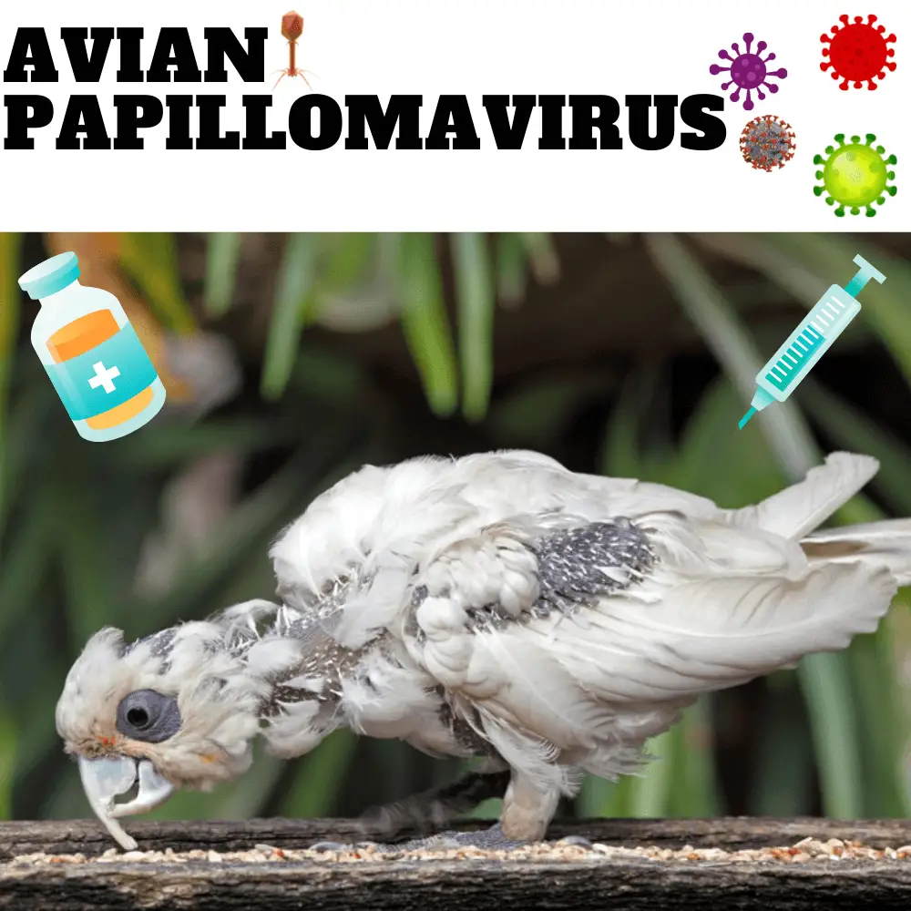
Avian papillomavirus: Here is a term which could frighten more than one… but do not worry about it! I will undoubtedly answer the first question that will come to your mind: no avian papillomas are not contagious to humans! You will not have to get rid of your bird ( or worse yet have it euthanized ) if it ever ( and I mean if ever, because avian papillomas are very rare ) is affected by papillomavirus or by papillomatosis ( definition of this term later in the text ). Second, you should know that if you have a bird with lesions, it will rarely need to be treated ( I will also explain why below ).
Avian papillomavirus
The virus
Family: Papovaviridae, a family which itself divides into two sub-families: Papillomavirinae ( which will be discussed in this text ) and Polyomavirinae ( polyomaviruses, viruses which are also well known in our parrots ).
Viruses of the Papillomavirinae subfamily are generally associated with the formation of benign skin tumors commonly referred to as papillomas.
The virus is highly specific to a single host, meaning that it is not easily transmitted from one species to another. Birds kept in close contact with other infected parrots very often remain healthy.
The lesions are usually proliferative ( they tend to multiply ) and rarely necrotic ( the word “neurotic” is the adjective of the word “necrosis” which means the death of living tissue ).
Avian papillomas are rather rare in psittacines. Some hypotheses could explain this rarity:
- many suspicious lesions are in fact not caused by a virus;
- lesions are caused by a virus, but the virus is not very transmissible;
- lesions are caused by a virus, but the majority of affected birds develop subclinical infections and immunity to the disease.
The incubation period is unknown, so it is very difficult to determine when, where, and how the bird was exposed to the virus.
Avian papillomavirus
Important points to remember
To date, all attempts to prove that the papillomatous masses found in the cloaca of birds were caused by papillomavirus have been unsuccessful. In the case of lesions found on the skin of finches and African gray finches, they are indeed caused by a papillomavirus, but they are the only ones. In birds, lesions resembling true papillomas were observed in several places throughout the alimentary tract. They are commonly found in the oral cavity and at the level of the cloaca. What causes these lesions remains unclear. To differentiate between true papillomas ( those caused by papillomavirus) and false papillomas, we will use the term papillomatosis which will refer to false papillomas.
First appearance in the bird
The first time that a virus of the Papovaviridae family was identified in a bird was in 1959 in finches. The virus was characterized by proliferative masses on the skin of the paws and feet. In African grays, persistent skin masses on the head and face have also been shown to be caused by papillomavirus.
Clinical signs
In general, parrots with papillomatous lesions show no clinical signs. In addition, the blood test remains normal, except in cases where
- internal lesions become necrotic;
- the food ingested remains trapped on lesions along the digestive tract, which induces an inflammatory response;
- the lesions are infected with bacteria or fungus.
Problems, therefore, arise when a lesion interferes with the ingestion and/or digestion of food or even when a lesion interferes with defecation.
When the lesions are found at the level of the cloaca, we can note:
- tenesmus ( continual almost unnecessary urge to have a bowel movement causing pain in the anal area );
- foul-smelling putrid feces or softer feces ( soiled feathers around the cloaca ;
- infertility ( depending on the location of the lesions and their severity );
- recurrent enteritis;
- recurrent prolapse.
When the lesions are found in the oral cavity or the esophagus, we can note:
- halitosis ( bad breath );
- dysphagia ( difficulty eating );
- dyspnea ( difficulty in breathing );
- wheezing.
When the lesions are found in the upper intestinal tract, we can note:
- anorexia;
- vomiting;
- chronic weight loss.
Avian papillomavirus
As for lesions found in the proventriculus, ventricle, and crop, the clinical signs can closely resemble PDD ( proventricular dilatation disease ).
It seems that parrots with papillomatosis also commonly have pancreatic or liver cancer. This could suggest that the cloacal or oral lesions could be caused by a virus that has the ability to transform infected cells.
Diagnostic
To diagnose papillomas, a histological evaluation ( study of tissues ) of the suspicious lesion taken by biopsy is necessary. In addition, it will be necessary to examine everything under an electron microscope to demonstrate the viral particles of 45 to 50 nm that are commonly found in inclusion bodies ( particles foreign to the cell ) in the nucleus of cells.
Visualization of papillomatous lesions at the level of the cloaca: a cotton swab soaked in physiological saline is used to evert the cloacal mucosa and a few drops of vinegar are applied. If the lesion turns white, it is most likely papillomatosis. Lesions in the cloaca are friable, reddish, pinkish, or whitish in appearance, and they tend to bleed profusely when handled or damaged.
Visualization of lesions in the digestive tract: it is possible to take an X-ray with a contrast medium ( that is to say that a coloring product is sent into the digestive tract to be able to identify the lesions on the X-ray ) or an endoscopy ( device equipped with a camera that can be inserted into the digestive tract to visualize the lesions ). Using the endoscope, it is possible to take a lesion specimen ( biopsy ).
Avian papillomavirus

Treatment for true papillomas
In general, true papillomas that are found on the skin of parrots do not require treatment. However, a damaged lesion could cause a secondary infection and should therefore be treated. Another possibility would be an obstacle to the natural movement of the bird by a lesion or even a lesion that would cause difficulty in gripping the food or in chewing the latter.
Mild lesions should be monitored. If changes occur and the lesions progress severely, it may be necessary to have them surgically excised. When this is the case, the lesions must be excised by radiosurgery ( destruction of tissue by irradiation which is done using a stereotaxic device to target the region to be reached ) to make the bird more comfortable and prevent injuries. secondary infections.
Treatment for papillomatosis
Papillomatous lesions may regress spontaneously and remain undetectable for a period ranging from 2 to 18 months. Then they can reappear. Lesions found in the oral cavity are often localized and therefore easy to excise and often non-recurrent. On the other hand, those at the level of the cloaca are often diffuse and therefore difficult to excise, and they tend to be recurrent. When it is necessary to excise papillomatous lesions, we can also use cryotherapy (the therapeutic use of intense cold to destroy tissue ), radiosurgery ( as mentioned above) or laser surgery. The use of silver nitrate is also possible to “burn” the papillomatous lesions at the level of the cloaca. However, with this type of procedure, the treatment should be repeated at 2-week intervals until the lesions have all resolved.
Prevention
Until we have more information on the etiology of papillomatous lesions, it would be advisable to isolate the infected bird from other birds.
Malnutrition, as well as hypovitaminosis A (vitamin A deficiency), could be predisposing factors. Good nutrition is PRIMORDIAL for the good health of your parrot; not only in this case but at all times. For more information on diet, I refer you to the chronicles: diet.
Bird Budgie Papillomavirus
SOURCE:Ross Perry
Reference
Branson W. Ritchie, Avian Viruses, 1995 Wingers Publishing, Inc.
