


A bit of history… It was in 1975 that Psittacine Beak and Feather Disease PBFD was first identified and formally described. It has since been recognized as the most important pathology in Australian Psittacidae.
Veterinarians around the world have sought to explain the disease by multiple causes. Some blamed sunflower seeds, while others mentioned inbreeding.
The Murdoch University research cell demonstrated that PBFD was caused by a new type of virus, the characteristics of which have since been established by researchers at the University of Georgia. Recent research has concluded that the disease is widespread among the wild population of cockatoos and other psittacines.
Beak disease causes
PBFD is caused by a relatively simple virus that infects and kills feather and beak cells. The virus also attacks the cells of the immune system, which it destroys. Thus many birds with PBFD succumb to secondary infections, bacterial or otherwise. PBFD circovirus is the smallest known virus capable of causing disease. This circovirus only causes problems in psittacines and as far as we know, no other species of bird or animal is susceptible to it. A disease similar to PBFD has recently been detected in doves: it is probably caused by a similar but antigenically different circovirus.
Psittacine Beak and Feather Disease
SOURCE:Evita Glez
Beak and feather disease symptoms
A bird with PBFD has characteristic features and most of the time, a simple clinical examination by a veterinarian can establish the diagnosis. PBFD usually affects young psittacines, but birds of all ages can succumb to the disease. PBFD in its chronic form is insidious in its development and progression, and dystrophic feathers replace normal ones as they molt progress.
Thus, a bird with PBFD may gradually lose its plumage, without any other symptoms. In cockatoos, the small powder feathers are often the first affected. Feathers affected by the PBFD virus are fragile or develop an abnormally thick outer covering.
Destruction of powder feathers results in areas of bare skin, and reduced powder production results in dull plumage and shiny beaks. Feather abnormalities are different depending on when the bird was in the molt cycle when the disease occurred. Abnormal feathers are usually short and have one or more of the following characteristics: defect in the lines, feather sheath too thick or skimpy; drop of blood visible in the quill of the feather.
The beak may also have abnormal growth or structure. In Euphemidae, apparently normal feathers that fall or are plucked effortlessly may be the only clinical sign. In birds with green plumage, the only clinical sign may be the appearance of yellow feathers, which otherwise appear normal. This discoloration is likely the result of microscopic changes in the structure of the feather.
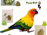
Other diseases often come on top of the PBFD: bacterial, fungal, and viral infections. Most birds with chronic illnesses eventually have difficulty feeding, lose weight and die. Severely affected birds often have mucous or green diarrhea, signs frequently diagnosed as secondary, bacterial, or chlamydial infections. The virus can also cause severe hepatitis, especially in cockatoos. The birds can then die of hepatitis without showing damage to the feathers.
Other diseases often come on top of the PBFD: bacterial, fungal, and viral infections. Most birds with chronic illnesses eventually have difficulty feeding, lose weight and die. Severely affected birds often have mucous or green diarrhea, signs frequently diagnosed as secondary, bacterial, or chlamydial infections. The virus can also cause severe hepatitis, especially in cockatoos. The birds can then die of hepatitis without showing damage to the feathers. Other diseases often come on top of the PBFD: bacterial, fungal, and viral infections.
Most birds with chronic illnesses eventually have difficulty feeding, lose weight and die. Severely affected birds often have mucous or green diarrhea, signs frequently diagnosed as secondary, bacterial, or chlamydial infections. The virus can also cause severe hepatitis, especially in cockatoos. The birds can then die of hepatitis without showing damage to the feathers. Severely affected birds often have mucous or green diarrhea, signs frequently diagnosed as secondary, bacterial, or chlamydial infections. The virus can also cause severe hepatitis, especially in cockatoos. The birds can then die of hepatitis without showing damage to the feathers. Severely affected birds often have mucous or green diarrhea, signs frequently diagnosed as secondary, bacterial, or chlamydial infections. The virus can also cause severe hepatitis, especially in cockatoos. The birds can then die of hepatitis without showing damage to the feathers.
Diagnostic
Well-declared PBFD is not difficult to diagnose. Hard-to-diagnose cases are birds showing only subtle signs, due to age or immunity. Histological examination of feather follicles is routinely done to confirm clinical symptoms, but is not suitable for diagnosing infections in incubation Circovirus can be detected on affected feathers by haemagglutination (HA) analysis and HI antibodies can be identified in blood, serum, plasma, or egg yolk. HA virus detection is currently the best method for identifying circovirus in feathers, liver, and droppings. It can be performed on growing feathers, or on “dry” feathers.
The analysis on feathers is preferred to that on droppings because birds very affected by PBFD do not excrete high concentrations of virus in their droppings and certain birds suffering from the chronic form only excrete it intermittently. Serology is useful for detecting flocks of birds infected with PBFD and for demonstrating the presence of antibodies in individuals. The presence of antibodies means that the bird has been exposed to circovirus and a high titer of HI antibodies in an adult bird suggests that the latter does not suffer from chronic PBFD.
Nesting birds with incubating infection or severe disease may show low and declining antibody titers. Please note that the test cannot detect passively transmitted maternal antibodies. Blood can be collected directly onto filter paper and only a few drops are needed, so even small parrots can be easily tested. The paper can dry out and transported to the laboratory does not require a refrigeration installation.
The blood test detects the antibodies developed against the virus. Be careful again, because the AH and IH analyzes cannot identify young birds incubating the disease! This is why it is recommended to perform a second test 60 days after the first, regardless of the result.
The paper can dry out and transported to the laboratory does not require a refrigeration installation. The blood test detects the antibodies developed against the virus. Be careful again, because the AH and IH analyzes cannot identify young birds incubating the disease! This is why it is recommended to perform a second test 60 days after the first, regardless of the result.
The paper can dry out and transported to the laboratory does not require a refrigeration installation. The blood test detects the antibodies developed against the virus. Be careful again, because the AH and IH analyzes cannot identify young birds incubating the disease! This is why it is recommended to perform a second test 60 days after the first, regardless of the result.
Incubation and vital prognosis
The incubation period for PBFD may be as short as 21 days, but the dose of virus, age of the bird, level of feather development, and lack of immunity must also be taken into account. The target organ is the epidermis and manifestation of the disease on the feathers requires molting.
Therefore, birds that contract the disease after their molt may not develop clinical signs until their next molt, which can last 6 months or more! Most birds that die of PBFD are less than 2 years old. However, all age groups should be considered susceptible to circovirus. Chronic exposure to high virus concentrations and/or stress are likely to be the conditions for infection and seroconversion of adult birds.
Prognosis
Spontaneous recovery after well-declared PBFD has been observed in several species, including budgies, lorikeets, and lovebirds. Severely affected birds can also recover. However, the majority of birds suffering from the chronic form of the disease do not recover.
PBFD in wild birds
The first outbreak of PBFD among wild birds probably occurred in 1888 in the Adelaide Hills, among a wild population of red-rumped parakeets (Psephotus haematonotus). Cases of PBFD have been confirmed in wild populations of Galah, Sulphur-crested Cockatoo, Bare-eyed Cockatoo, Rainbow Lorikeets, Orange-bellied Parakeet, Omnicolor, Rose-ringed Parakeet, Major Mitchell’s Cockatoo, Gang-gang, king parakeets, Latham (or swift) parakeets, budgies, red-rumped parakeets, funereal cockatoos, blue-capped parakeets and many more. Groups of wild cockatoos could present a proportion of 20% of infected birds and a seroprevalence of 60-80%. The infection is maintained in the population through sick birds. Outbreaks can occur in groups of wild or aviary birds. The virus is probably transmitted horizontally, but carrier birds could contribute to vertical transmission. The virus is likely to remain pathogenic in contaminated nests for several months or years.
PBFD in aviary birds
Groups of aviary birds with cases of PBFD generally display a high seroprevalence. In these cases, the birds affected by PBFD are often the offspring of hens with low or undetectable serious antibody levels.
How to prevent and control PBFD?
It is recommended that breeders keep newly purchased birds isolated from healthy birds, and even breed them in quarantine Disinfectants (such as glutaraldehyde) capable of inactivating environmentally resistant viruses such as parvoviruses are recommended to disinfect utensils, cages, and contaminated parts. PBFD is difficult to quarantine. Carrier birds may appear clinically healthy but produce diseased young. Hence the need to breed quarantined birds.
Vaccination (Inactivated vaccine)
A killed virus vaccine was recently developed in Australia. Administered to healthy birds, the vaccine stimulates immunity to circovirus. This vaccine is not a treatment for birds that already have PBFD as it is not curative. It can even worsen the development of the disease. It is important to vaccinate pet birds when they are young, in fact as early as 14 days old. All vaccinated birds should be boosted one month after the first injection. Thereafter, the bird will need to be examined every 6 months, until it is 3 years old. Breeders should also be vaccinated one month before the breeding season. Please note that this vaccine is not available in France. Some veterinarians dispute its effectiveness.
Psittacine Beak and Feather Disease
A virus is known for a long time
This disease was first studied in 1975 on Australian parrots. It is caused by a circovirus, which mainly affects Australian parrots. It evolves over several years.
Alteration of feathers and beak
The virus prevents the normal production of keratin by the body, which is the protein that goes into the formation of horny productions: feathers, beak, and scales of the legs.
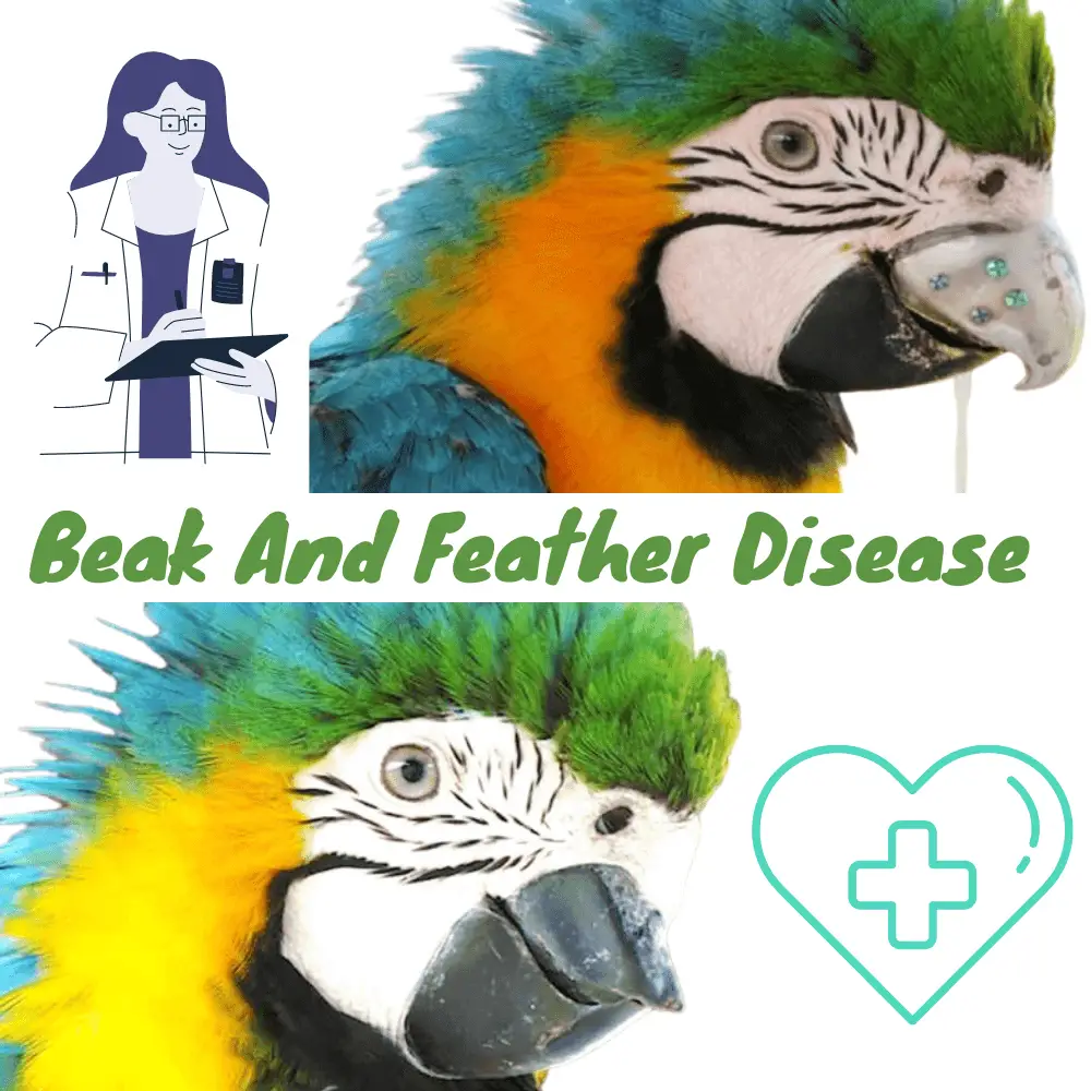
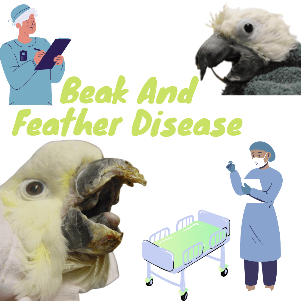
Symptoms start with a deformation of a few feathers after the molt, the new shoots not developing properly. The feathers remain hooded in their sleeves and sometimes have small spots of blood at their base. Little by little, the bird moves around until it becomes completely naked. The cockatoo beak becomes shiny because the keratin powder which is normally present on its surface is no longer properly synthesized (see photo 1: cockatoo with PBFD).
The scales of the legs deform and the beak loses its rigidity until it softens completely and sometimes falls in extreme cases.
In the wild, the bird, unable to fly and feed, dies quickly. In captivity, he can live much longer (several years) with the virus, thanks to the assistance of his master.
Immunodeficiency
This disease also causes an immunodeficiency (attack of the immune defenses) which weakens the bird even more by allowing the appearance of all kinds of other opportunistic infections.
A confusing disease that evolves differently depending on the parrot species
Among the affected birds, many remain healthy carriers of the virus without developing the disease. Despite everything, they are contagious.
The parrots most susceptible to this disease are Australian parrots, such as all varieties of cockatoo or Eclectus. Damage in a cockatoo is very serious because it generally leads to the death of the animal.
South American parrots (Macaws, Amazons Parrot, Pionus, etc.) are much more resistant and generally eliminate the virus spontaneously after a few months.
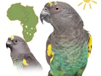
The sensitivity of the African Grey Parrot seems intermediate: very few adults affected to develop the disease, most remain healthy carriers. On the other hand, this virus is devastating for young Grays between weaning and 6 months, because they can die suddenly in two to three days without any treatment is possible.
Parakeets (wavy, cockatiel, lovebirds …) are also sensitive, but most often make attenuated forms (see photo 2, budgerigar with PBFD). They can however play the role of virus reservoir and transmit it to parrots.
Easy diagnosis
There are PCR tests that can detect the DNA of the virus in the bird’s blood or from the tip of the pen. These tests have been known for a long time and their results are reliable.
A positive test does not mean the same thing depending on the species of parrot:
A positive cockatoo unfortunately has every chance of developing the disease.
A positive adult African parrot (Gray, Senegal Parrot, etc.) that does not show symptoms remains generally positive, rarely develops the disease, but remains contagious for its congeners and young. However, it is possible that it will clear the virus spontaneously over time.
A positive South American parrot (Macaw, Amazon, etc.) only very rarely develops the disease. It is likely that it will spontaneously clear the virus after a few months. It is, therefore, useful to repeat the PCR test after 4 to 6 months after a positive test to check if the bird is still a carrier.